.png)

UCLA NICU Common Conditions
Neurology
Intraventricular Hemorrhage (IVH):
-
Pathophysiology: Intrinsically fragile and highly vascularized germinal matrix layer near the ventricles which is very vulnerable to hypoxia, blood pressure changes, respiratory failure with fluctuations in CO2, which can cause ischemic damage and changes to cerebral blood flow. The germinal matrix starts to significantly involute at 32 weeks and has nearly disappeared by 36 weeks, which is why late preterm infants are not at high risk for a germinal matrix bleed and the most at risk population is VLBWs and <32 week infants. Risk for IVH increases with lower gestational age and lower BW. However, term infants can still get IVH and ICH (intracranial hemorrhage) even though they are less at risk for brain bleeds.
-
Grading of IVH:
-
Grade I: Bleeding confined to germinal matrix
-
Grade II: Intraventricular bleeding (bleeding into < 50% of ventricular area with no ventriculomegaly)
-
Grade III: Intraventricular bleeding (bleeding into ventricular area with ventriculomegaly)
-
Grade IV: Intraparenchymal echodensity (IPE) represents periventricular hemorrhagic infarction
-
-
Prevention: Avoid unnecessary and frequent handling of infants in first 72 hours of life as part of neuroprotection bundle (do Q6H cares and keep baby in midline position). Target normal body temperature, normal blood pressure, normal CO2 level.
-
Screening: Head US ~DOL7 for all infants born at <32wks GA or <1500g. Can consider screening older/bigger infants who have had significant resuscitation and/or postnatal instability. Can obtain screening US earlier in any age based on. clinical concern and if results may guide clinical decision making (ex: wanting to start indomethacin (increases bleeding risk) for PDA closure
-
Symptoms: Seizures, persistent metabolic acidosis, decreased perfusion, hypoxemia, hyperglycemia, apnea, hyperkalemia, bulging fontanelle lethargy, or rapid unexplained drop in hematocrit.
-
Follow-up: If first HUS is abnormal, repeat Q1-2wks. Follow daily head circumference if Grade 3-4 IVH (or enlarged ventricles) are present and consider biweekly HUS with neurosurgery input. If no IVH, repeat head ultrasound at age 4-6 weeks for detection of cystic PVL.
-
Consider brain MRI before discharge for those with more severe IVH.
-
If there is significant or increasing hydrocephalus on HUS, neurosurgery will need to be consulted for potential drain placement.
Cephalohematoma:
-
Bleeding of small subperiosteal blood vessels which causes separation of the periosteum from the calvarium.
-
Well circumscribed
-
Does not extend across suture lines
-
Can range from soft to more tense depending on how large the bleeding is. Larger ones will be more protuberant given inability to cross suture lines and can have a golf ball appearance.
-
Becomes more noticeable hours to days after birth
-
Can take weeks to months to resorb
Subgaleal Hemorrhage:
-
Bleeding of emissary veins (connections between the dural sinuses and the scalp veins) between the epicranial aponeurosis of the scalp and the periosteum.
-
Crosses sutures lines
-
Large boggy swelling that can extend into the nape of the neck
-
Extensive bleeding can cause facial edema, proptosis and displacement of the ears
-
Infants with possible subgaleal should get serial hematocrits and close monitoring of head circumference and hemodynamic vital signs.
-
Neonates can lose 50-70% of their circulating blood volume in the subgaleal space and thus are at high risk for mortality and need to be monitored very closely and have a high index of suspicion for this underlying process.
-
With progressive blood loss, infants will develop signs of hypovolemic shock. Normal HR and BP can be falsely reassuring in an infant with a subgaleal bleed and does not exclude the possibility of hemorrhage.
-
Can be associated with DIC; check coagulation factors and replete as necessary.
-
US and CT can be used to help differentiate a subgaleal bleed.
-
Most often associated with vacuum or forceps-assisted delivery, although can occur spontaneously.
Caput Succedaneum:
-
Serosanguinous fluid accumulates in the subcutaneous layer of the scalp.
-
May cross sutures lines and can cause confusion with subgaleal bleeds, however is usually not as expansive as a subgaleal and will resolve in <24 hours.
Retinopathy of Prematurity (ROP):
-
Pathophysiology: Premature infants have an immature, incompletely vascularized retina which leads to the development of terminal vascular shunts and neovascularization that, in its most severe form, causes tractional retinal detachment and blindness.
-
Grading: Zone (how far abnormal blood vessels extend over retina), Stage (degree of tortuosity of blood vessels), PLUS disease (significant tortuosity of vessels, high risk of blindness, often treated with VEGF inhibitors and then laser therapy). High zone, low stage = good prognosis vs low zone, high stage = bad prognosis.
-
Prevention: Limit over or under supplementation of oxygen, screen at risk populations and closely monitor to prevent disease progression and retinal detachment.
-
Screening: Infants of birth weight < 1500 gm or <30 weeks GA receive ophthalmology exam at 1 month of 31 weeks corrected GA (whichever is later) to evaluate for ROP. Can also consider screening infants who do not meet this criteria if they have unstable courses (severe hypoxemia, hypotension, etc).
-
Follow-up: Per Ophtho
Hearing Screens
-
All infants should be screened prior to discharge and again at 90 DOL.
-
California hearing screening guidance can be found here.
-
Evoked OAE: Otoacoustic emissions
-
Can be performed when infant is awake
-
Simpler testing technique
-
Detects outer/middle ear and/or cochlear problems
-
A sensitive microphone/probe is placed in the ear and detects signals in response to an auditory stimulus.
-
Does not identify auditory nerve or auditory brainstem dysfunction or mild forms of hearing loss. Because of this, infants with a normal OAE may fail an ABR.
-
Vernix can cause higher false positive (fail) rate. Because of this, infants with a failed OAE may have a normal ABR.
-
-
Automated ABR: Auditory brainstem response
-
This is the hearing test we provide in the UCLA NICU
-
Best performed when infant is asleep
-
Requires more operator skill and electrodes are placed on baby’s scalp
-
Cochlea, auditory nerve, and brainstem dysfunction are detected
-
Difficult to perform on older infants without sedation
-
Even if ABR or OAE test results are normal, hearing cannot be definitively considered normal until a child is mature enough for a reliable behavioral audiogram to be obtained.
-
Hypoxic Ischemic Encephalopathy (HIE):
-
Pathophysiology: Perinatal event that causes impaired cerebral blood flow and oxygen delivery to the brain with resulting primary and secondary energy failure and neonatal encephalopathy. Often associated with a sentinel event: abnormal fetal heart tracing such as variables or late decelerations, placental abruption, uterine rupture, cord prolapse, maternal hypotension, and more.
-
Criteria for cooling at UCLA:
-
Cooling must begin within 6 hours of birth to be of benefit
-
Infant at > 36 weeks GA *can consider cooling infants even younger after weighing risk/benefits and in shared decision making with families
-
Apgar score < 5 at 10 minutes after birth
-
Continued need for resuscitation 10 minutes after birth
-
Acidosis defined as either umbilical cord pH or any arterial pH within 60 minutes of birth < 7-7.1, base deficit > 10-16 in umbilical cord blood sample or any blood sample within 60 minutes of life (arterial or venous)
-
Infant with moderate to severe encephalopathy (see table below). Mild encephalopathy/borderline infants can be cooled on a case by case basis after shared-decision making with family on risks/benefits and concern of the primary team.
-
-
Cool for 72 hours and then rewarm. Perform MRI/MRS (with HIE protocol) after rewarming. Patterns of injury that can be seen in HIE are: thalamus and basal ganglia and posterior limbs of the internal capsules; watershed injury involving the cortical and subcortical white matter; focal or multifocal minimal white matter injury; extensive whole brain injury. The MRS can tell us about metabolite peaks that are seen in areas where brain tissue has seen ischemic injury and has had to undergo anaerobic metabolism.
-
EEG can be discontinued 24 hours AFTER rewarming
-
Complications: DIC, metabolic acidosis, electrolyte derangements, seizures, tachyarrhythmias, bradycardia, apnea, pulmonary hypertension
Respiratory
Indications for mechanical ventilation:
-
pH < 7.25, with PaCO2 > 50 mmHg
-
Requiring FiO2 > 0.35 - 0.4 to achieve target SpO2 goal
-
Apneic episodes or neurological concerns causing respiratory compromise (status epilepticus, CNS malformation, HIE, medication side effects (ex: PGE))
Normal range of arterial blood gas values for term and preterm infants at normal body temperature and assuming normal blood Hb content (Source: Gomella 8th edition)
-
Venous blood gas: pH values are slightly lower, PCO2 values are slightly higher, and PO2 is of no value in assessing oxygenation.
-
Capillary blood gas: Heel should be warmed before sampling. pH usually slightly lower, PCO2 values are slightly higher, but this may vary based on sampling technique and perfusion.
Invasive modes of respiratory support:
-
Conventional Mechanical Ventilation (CMV): In our NICU, we use SIMV/PC (as opposed to AC modes and VG modes). Starting settings: Rate: 30-40, deltaP 10-20, PEEP 5-6, PS: 6-8. When on a pressure mode, the dynamic variable will be your tidal volume. To prevent volutrauma, can target TV of 4-6 cc/kg as a minimum to achieve adequate ventilation.
-
Synchronous intermittent mandatory ventilation (SIMV): A set rate will give you full “mandatory” ventilator breaths (either your target PIP or target volume). Breaths that baby takes “over” your set rate will be pressure supported breaths (generally, will be a smaller PIP/effective TV than your regular ventilator breaths). Pressure supported breaths appear as orange/red on the ventilator.
-
Assist control (AC): In AC modes, every breath that is triggered by the baby will be a full “ventilator breath.” There are no pressure supported breaths, thus there is no variation in the target pressure or volume that is given to the baby each breath, depending on the mode that is used (PC or VG).
-
Pressure control (PC): The target PIP is set, the dynamic variable is the tidal volume. As lung compliance improves, the tidal volumes will increase for the same delivered pressure (ex: post-surfactant administration). As lung compliance worsens, tidal volumes will decrease for the same delivered pressure (chronic lung disease). Can be SIMV/PC or AC/PC.
-
Volume guarantee (VG): The target volume is set, the dynamic variable is the pressure delivered to reach the target volume. As lung compliance improves, less pressure is needed to achieve the target volume (ex: post-surfactant administration). As lung compliance worsens, delivered pressures will increase to achieve the same target volume (chronic lung disease). Can be SIMV/VG or AC/VG.
-
-
High Frequency Oscillator Ventilation (HFOV): A piston pump generates pressure in the ventilator circuit. Low-tidal volumes and high frequency oscillations around a target/constant mean airway pressure (in contrast to the inflate/deflate cycle caused by conventional ventilator).
-
-
Consider HFOV when needing high PIPs to achieve adequate ventilation on CV, poor oxygenation on CV, air leak syndromes, pulmonary hypoplasia and more.
-
Set Hz (~ RR) = 12-14 for preterm infants and 10 for term infants, Mean Airway Pressure (MAP) = 2-4 above the settings on the CMV, Amplitude (~TV). Generally, your AMP should be no more than 2-3x the MAP and should be increased to achieve good chest wiggle.
-
Monitor lung fields closely for hyperinflation and decreased cardiac silhouette which can lead to decreased cardiac output.
-
To decrease your CO2: Increase Amp or decrease Hz. Generally, Amp is maximized before decreasing your Hz. Decreasing Hz will cause a larger change to your CO2 than micro changes to your Amp. To increase oxygenation, increase the MAP. Downside: can have atelectasis due to smaller tidal volumes and inability to give “sigh” breaths (aka conventional ventilator breaths), decreased cardiac output, overexpansion
-
-
NAVA: Can be invasive or non-invasive. An esophageal probe gives you feedback on diaphragmatic contractility and delivers pressures based on how hard a baby is working to breathe.
-
-
Generally, start at level of 2, Apnea time of 2 seconds. Set up desired back up pressures and peep. Goal EDI peak < 10-15 and goal EDI minimum < 5.
-
If EDI peaks are high (red graph), increase your NAVA level. When your EDI peak is within a goal range or lower, the NAVA level may be weaned by 0.2. Generally, wean to a minimum of NAVA level of 1.0, but can decrease even further as well (0.8, etc) before coming off the modality.
-
Delivered PIP will vary with each breath and depend on your EDI peaks/minimum (Delivered PIP = Nava Level x (EDI peak - EDI min) + PEEP). iNAVA is more synchronous then conventional ventilators because it detects a baby’s inspiratory breath from the neural diaphragmatic excitation stage vs. conventional ventilators use a flow trigger to detect a baby once they are already inspiring (see diagram below).
-
Non-invasive modes of respiratory support:
-
Nasal cannula: ¼ -2L
-
High flow: NC: 2-6L
-
Vapotherm/’Precision Flow’: Similar to highflow in delivery, air is more humidified and provides more laminar and higher velocity flow (at the same flow rate when compared to HFNC). Use for infants who are more premature.
-
CPAP: Set PEEP 5-6 - bubble CPAP or CPAP via the ventilator and various interfaces for delivery of PEEP: RAM cannula vs. Fisher & Paykel mask. Discuss with RT and bedside nurse what is most appropriate for patient. Preterm infants generally do better on bubble CPAP.
-
Fisher Paykel
-
-
-
RAM cannula
-
-
NIPPV or NIMV: Rate 20-40, delta P 15-20, PEEP 5-6. Best mode to prevent extubation failure and/or for infants who are having apneic events.
-
NIV-NAVA: See above
-
For preterm infants on non-invasive support, keep on bubble CPAP if tolerated until about 32 weeks CGA. It’s best to wean PEEP slowly to allow for optimal growth and prevent atelectasis and reduce rates of BPD.
Apnea of prematurity
-
Centrally mediated, common before 34wks GA, caused by physiologic immaturity of respiratory control. Cessation of respiration for 20 seconds or < 20 seconds if associated with hypoxemia and/or bradycardia. Apnea that is < 10 seconds is considered “significant” if it is associated with a decrease in SpO2 to 80% or less or bradycardia to < 80.
-
Treatment:
-
Caffeine (load 20mg/kg; 5-10mg/kg daily thereafter). Side effects: tachycardia, restlessness, GERD
-
IV to PO conversion is 1:1
-
NC – respiratory stimulation and support
-
Can consider discontinuing once >34 weeks CGA and on minimal or no respiratory support with minimal or no apneic events
-
Respiratory Distress Sydrome (RDS)
-
Most common respiratory disorder in preterm infants, typically affects infants35 weeks GA with an additional risk factor (infant of diabetic mother for example) may also have clinically significant RDS.
-
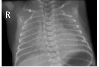
Clinical diagnosis: tachypnea, retractions, grunting, nasal flaring. Primary cause is inadequate pulmonary surfactant; which leads to decreased compliance and a tendency to atelectasis. Typical CXR shows low lung volumes and a bilateral reticular granular pattern (ground glass appearance) with superimposed air bronchograms.
-
Management: antenatal glucocorticoids, CPAP and PEEP and surfactant replacement therapy, time (endogenous surfactant increases over first few days). Surfactant given via ETT or non-invasively using “LISA”. Complication is pulmonary hemorrhage and pneumothorax 2/2 rapidly increased lung compliance.
-
Best to give surfactant as soon as possible for premature infants that show signs of surfactant deficiency: significant respiratory distress, FiO2 requirement >30%, etc. as seen on Lung injury prevention clinical pathway.
Transient Tachypnea of the Newborn (TTN)
-
TTN is a benign disease of near-term and term infants who have respiratory distress shortly after delivery that usually resolves within hours to 3 days. Risk factors include c/s delivery or precipitous VD delivery. Incidence is ~1-2% of all newborns. Delayed resorption of fetal lung fluid is thought to be the cause.
-
Signs include tachypnea and retractions, which resolve. Classic CXR finding "fluid in the fissure" (minor fissure), hyperexpansion of lungs, prominent perihilar streaking (engorgement of periarterial lymphatics).
-
CPAP or nasal cannula are often sufficient, however some infants need NIMV. PO feeding is OK if resp rate <60 and <2L HFNC without significant work of breathing, otherwise OG feed. If significant work of breathing, patient should no be allowed to orally feed.
-
Rule out: pneumonia, CHD, RDS, cerebral hyperventilation, metabolic disorders, polycythemia and hyperviscosity. TTN is self-limited and infants generally rapidly wean off of respiratory support.
Meconium Aspiration Syndrome (MAS)
-
Meconium stained fluid is evidence that the fetus passed stool in utero. Meconium may be aspirated if the fetus gasps.
-
Risk factors for MAS include: postterm pregnancy, preeclampsia, maternal hypertension, maternal diabetes mellitus, abnormal fetal heart rate.
-
CXR shows heterogeneous lung with areas of hyperexpansion and atelectasis and diffuse, patchy infiltrates.
-
Surfactant has been shown to be beneficial in these infants as meconium in the lungs inactivates endogenous surfactant.
-
Infants with MAS are at risk for pulmonary hypertension. May need HFOV and nitric oxide and even ECMO if significant PPHN.
Bronchopulmonary Dysplasia (BPD)/Chronic Lung Disease (CLD)
-
BPD is CLD that affects premature infants. Generally defined as requiring some form of respiratory support at 36 weeks postmenstrual age if born < 32 weeks GA or >28 d but < 56 d postmenstrual age or discharge to home, whichever comes first.
-
BPD can be classified as grades 1, 2, or 3 depending on the respiratory support they are on at 36 weeks PMA. BPD is caused by a combination of prenatal and postnatal factors in addition to premature delivery. Premature delivery causes arrest of the normal process of alveolarization and lung development. Aberrant repair mechanisms to the pre and postnatal insults over time results in inflammation, fibrosis, and heterogeneous lung injury that impairs gas exchange and leads to high resistance airways. This process co-occurs with abnormal microvascular development which also makes infants with BPD at risk for pulmonary hypertension. Chorioamnionitis, IUGR/FGR, and maternal smoking are examples of prenatal insults. Mechanical ventilation, sepsis and oxygen toxicity as examples of postnatal insults.
-
Lung inflammation is a huge component of BPD development, thus postnatal steroids can be considered for its anti-inflammatory effects. See Respiratory Care Guidelines for ELBW infants.
-
The physiology of BPD changes over time of acute vs established disease. An overview of “BPD settings” are shown below:
Cardiovascular
PDA (Patent Ductus Arteriosus)
The ductus arteriosus is patent in all newborns at the time of delivery. It is closed by 48h after birth in 100% of infants delivered at ≥40 wks gestation and by 72 h after birth in 90% of infants delivered at ≥30 wks gestation. A ductus open beyond 72 h can be considered to be a persistently patent ductus arteriosus. Around 30-40% of VLBWs have a PDA.
Signs/symptoms: wide pulse pressure, bounding pulses, palmar pulses, systolic/diastolic murmur, pink-tinged airway secretions, pulmonary hemorrhage, pulmonary overcirculation, feeding intolerance
Stepwise treatment if hemodynamically significant: signs of pulmonary overcirculation and ductal steal from systemic circulation.
Fluid restriction
Indomethacin Q12h x3 doses,Treatments may be repeated. In-between doses follow UOP (goal >1 cc/kg/hr), Cr (watch trend), Platelets (goal >100). If UOP is between 0.6 cc/kg/hr to 1 cc/kg/hr, 24 hour dosing interval should be used. Doses should be withheld if the patient has UOP < 0.6 cc/kg/hr. Because indomethacin decreases gastrointestinal blood flow, infants may be kept NPO until at least 24 - 48 h after the indomethacin therapy has been completed. Contraindications to indomethacin use include intracranial bleed, bleeding diathesis, thrombocytopenia and renal insufficiency. Should not be used at the same time as steroids due to increased risk of spontaneous intestinal perforation. Tylenol is preferred in these patients.
Dosing:
PNA at time of first dose PNA at time of first dose 2 to 7 days: IV: 0.2 mg/kg at 12- to 24-hour intervals.
PNA at time of first dose >7 days: IV: 0.25 mg/kg at 12- to 24-hour intervals.
If contraindications to Indomethacin, can use IV Tylenol 15 mg/kg Q6H x 3 days.
Surgical ligation (done by pediatric surgery) or device closure with piccolo (done by pediatric cardiology)
When, how and if one should close a PDA in a given infant is highly controversial
Types of Access
All central lines need weekly surveillance XR
All central lines (PD PICC, Neo PICC, UVC, UAC, Broviac) need heparin! If the fluid is going to run at 1 cc/hr or more (this applies to most fluids you will order in the NICU), heparin can be 0.5 U/mL. If the fluid (usually only UAC fluids) are going to run at 0.5 cc/hr maximum, heparin must be at 1 U/mL.
A NeoPICC and UAC always needs a continuous fluid running through it. A double lumen UVC has one line running a continuous infusion while the other line may be capped. Therefore, if you are stopping TPN and maintenance IVF but you are keep a UVC in for medications, you will have to order a carrier fluid (usually NS) of 1 cc/hr.
PALS (peripheral arterial line) also needs heparin, same ordering principles as a UAC. Should be run at 1 cc/hr or more to decrease change of losing the line.
Heparin flushes: Use the order set.
Double lumen UVC: 10U/1mL heparin Q6H (capped line) and 10U heparin Q24H (continuous infusion line)
NeoPICC: 10U/1mL Q24H
PD PICC: Old practice: 30U heparin Q24H New practice: Normal saline flush Q12H (minimum 1 mL)
Subclavian/IJ/Femoral Line: Old practice: 10U/1mL Q6H (capped line) or 10U/1mL (continuous infusion line) New practice: Normal saline flush Q12H (minimum 1 mL)
PIV: Max dextrose 12.5%
Central line: Max dextrose 30%
Note: Calcium is generally reserved only for central lines. Calcium should only be given peripherally on a case by case basis with explicit permission of the fellow or attending due to risk of extravasation and damage to surrounding tissue.
Note: For infants with a PIV, peripheral concentration (0.1 mEq/mL) of potassium (as delineated in the order itself) should be used to replete.
If an infant has a central line, use the central concentration (0.4 meq/mL)
PICC: needed if plan for prolonged TPN ( 2.5 kg and do not require continuous infusions (after the first 24 hours).
UVC: placed for premature or term infants expected to need prolonged IV nutrition or medications for critical illness. Can be used to infuse nutrition and for lab draws. Generally kept in for no more than 7-10 days (but up to 14) except in extreme circumstances where umbilical central access must be maintained due to critical illness and/or inability to obtain another source of access. Note: Low-lying UVCs are not considered central lines. Their osmolar concentration should be limited to UAC: placed for infants who need frequent and/or arterial lab draws (for arterial lactate and PaO2 monitoring) and/or blood pressure monitoring. Cannot be used to infuse nutrition. Generally kept in for no more than 7-10 days except in extreme circumstances where umbilical arterial access must be maintained due to critical illness and/or inability to obtain another source of access. Generally not kept in when advancing past trophic feeds or low volume feeds (20-40 cc/kg/day). Start with 1/2NaAcetate or 1/2NS to run at 0.5-1 cc/hr. Consider an infant’s acid base status when deciding which one to order. Generally, premature infants are started on 1/2NaAcetate because premature kidneys mimic a Type 2 bicarbonate wasting RTA and have metabolic acidosis. See Arterial Fluids Panel (NICU) Order
UAC length = 3xBW (kg) + 9
UVC length = 1.8xBW (kg) + 4.7 or UAC length divided by 2 + 1
The appropriate location of the UVC on XR is approximated based on its location on XR: should be the junction of the IVC and RA. POCUS is a great way to confirm the location of the UVC as XRs only provide an approximation. UVCs often migrate and may require adjustment over the early days after placement. A lateral film can also help delineate the location of the UVC further. Deep UVCs should be pulled back. If the UVC migrates into the liver, it should be pulled to a “low-lying” position (outside the inferior margin of the liver) as infusing parenteral nutrition directly into the liver is associated with hepatic injury. Complications of UVCs include: infection, pericardial perforation with pericardial effusion and tamponade, arrhythmia, hepatic injury (necrosis, hematoma), portal vein thrombosis, air embolism, catheter dislodgement and more.
The appropriate location of the UAC is approximated based on its location in relation to the thoracic vertebrae. The following pneumonic can be used to approximate an appropriate UAC position: “7 is heaven, 8 is great, 9 is fine, 10- do it again.” Thus, ~T8 is the ideal UAC position because it is safely above the celiac artery and avoids the renal arteries and superior mesenteric artery. High UACs are preferred (compared to low-lying UACs which are at the L3-L4 level) because they are associated with less lower extremity complications (ischemia, thrombosis). Complications of UACs include: infection, vessel perforation, thrombosis, ischemia of lower limbs and kidneys, hypertension and more.
The path of the UVC is: umbilical vein > ductus venosus > IVC/RA junction and is thus a shorter depth of insertion than the UAC.
The path of the UAC Is: umbilical artery > internal iliac artery > common iliac artery > descending aorta and is thus a longer depth than the UVC.
Ideal position: Shown in non-dashed lined
Low-lying: Shown in dashed line
Mean Arterial Blood Pressure (MAP)
Should be at least GA in the first day of life; goal is usually a minimum PMA in weeks
True hypotension is a clinical diagnosis based on your MAP and other markers of cardiac output and systemic perfusion: UOP, lactate, capillary refill, pulses
Here is one reference guide for MAP goals in premature infants
FEN/Growth
Fluids
See TPN section for details on fluid goals
Start with D10W on DOL 0 without electrolytes for term infants or starter TPN (D10 with AA) for premature infants. Add electrolytes to IV fluids or start PPN or TPN on DOL 1.
TPN
Needed in most infants born See TPN section for details about TPN contents and electrolyte goals.
Feeds
Considered “trophic” if 20-30 cc/kg/day or less. These low volume feeds are given to stimulate the gut and do not provide meaningful nutrition and thus are not included in the total fluid goal.
PO trialing starts at ~34wks PMA (do not have oromotor development to PO before this age) – consult OT around this time to assist
Caution advancing past trophic feeds when an infant has a UAC
For nutritional content of milk and formulas, click this link.
Fortification of Feeds:
See feeding protocol. Some premature infants may need a combination of EBM and 22 kcal formula (2-4) bottles prior to discharge for adequate growth. Some infants need to go home on even higher fortification to sustain adequate weight gain. See Individualized Discharge Feedings handout on the Box.
Because infants with certain congenital heart disease have higher risk of NEC, they follow a different feeding protocol, which can be found here.
Note that this feeding protocol only addresses the first two weeks of a cardiac infant’s life. The feeding plan beyond this time period should be individualized and depends upon an infant’s physiology, repair plan and risk factors for NEC.
Intolerance to Feeds
Signs: emesis, blood in stool, metabolic acidosis, onset of apneic events,distended, firm or discolored abdomen, “loose stools” or diarrhea, temperature instability, hyperglycemia
May be a sign of necrotizing enterocolitis (see below) or prematurity-related feeding intolerance; consider holding feeds, reducing volume, extending length of feeds, running continuous feeds, hydrolyzed feeds or breast milk only.
Gastric Residuals
Should not be routinely collected. Bedside nurse may report only if residuals return when venting for air.
Bilious residuals should be investigated further to make sure it is not bowel obstruction or another sinister pathology.
Growth
Goal average of 30g/day for term infants
Term infants should lose up to 8% of their BW and for preterm infants up to 10% of their BW. Infants have a higher total body water content with increasing prematurity (up to 90% in micropremies) and have more immature kidneys that leads to a very high urine output, which contributes to higher weight loss in the first two weeks of life compared to more mature infants.
All infants should regain BW by 2 wks or ideally sooner.
Use birth weight for ALL calculations until infants regains birth weight
Multivitamins
All infants need Vitamin D either through a multivitamin or vitamin D supplement.
Term infants need 400 IU (10 mcg) Vitamin D and should not receive multi-vitamin.
Preterm infants need 1 mL of multivitamin. This is added through TPN when on parenteral nutrition and is converted to an enteral supplement once tolerating full or close to full feeds. Generally split into 0.5 mL BID when infants are smaller to improve tolerance. Can be given as one mL a day of Poly-Vi-Sol (with or without Fe depending on infant’s Fe needs) when going home for ease of administration for caregiver.
Infants on full feeds fortified with Enfamil HMF to 24 kcal get enough Vitamin A, D, E, C, etc. from the fortifier and do not need to be on Poly-Vi-Sol, just 5-10 mcg Vit D (depending on volume of enteral feeds). However, once they are de-fortified, they should be switched back to Poly-Vi-Sol.
For a reference guide on how much of each vitamin is in the fortifier and what the goal vitamin doses are, click here and here.
1 mL of Poly-Vi-Sol with Fe has 11 mg Fe per dose and thus the Fe content in it cannot be titrated, so depending on an infant’s weight and Fe needs, you may or may not be able to send infants home on the combined Poly-Vi-Sol with Fe formulation. Infants who do not get combined Poly-Vi-Sol with Fe get separate prescriptions for Fe and Poly-Vi-Sol
Only infants taking at least 1 L of standard formula get enough Vitamin D to not need any Vitamin D supplementation.
Gastroenterology
NEC (Necrotizing Enterocolitis)
Ischemic and inflammatory necrosis of the bowel primarily affects premature neonates after the initiation of enteral feeds. Mortality of 10-50% depending on severity. Multifactorial etiology: prematurity, formula feeding, anemia, inflammation, bacterial colonization that results in intramural gas accumulation (pneumatosis intestinalis). NEC usually occurs at approximately 3-6 weeks of life; NEC occurs shortly after birth “in bigger babies” and much later (>8 weeks) in the “smallest” babies.
Signs & symptoms: feeding intolerance (bilious emesis/residuals), apnea, hyperglycemia/hypoglycemia, increased abdominal girth, abdominal distension, bluish/reddish discoloration of abdomen, abdominal tenderness, guaiac positive stools/bloody stools, increased or decreased WBC and decreased platelets, metabolic acidosis, hyponatremia/electrolyte abnormalities. See the Modified Bell’s Criteria below for NEC classification.
Work-up: KUB, cross table lateral film, looking for bowel dilation, thickening of bowel wall, fixed, dilated loop, portal venous gas, pneumatosis or free air.
Treatment: NPO, place replogle to low intermittent suction, sepsis work-up and start antibiotics. Antibiotic regimen must cover gram positive, gram negative and anaerobes. (Possible regimens include: amp/gent/flagyl or zosyn. Vancomycin can be added in severe illness or hx of MRSA colonization), check serial films and pertinent labs (CBC, CRP, electrolytes). Don't forget to adequately fluid resuscitate these babies. Consider Peds Surg consult. Follow-up films should include KUB and cross table lateral looking for perforation or resolution. Treatment protocol can be found in the Box under Antibiotic Stewardship.
Hematology
Indirect Hyperbilirubinemia
Indications for phototherapy: See stanford premie bili tool for infants < 35 wga and >48 HOL. For infants < 27 weeks, use 5 as a light level or do 5 x BW to approximate the light level. For infants > 35 wga, use standard bili tool.
Order cord blood work up (blood type, Rh and Direct Coombs), check mom’s blood type. If Coombs+, check retic. (retic >6 is a sign of hemolysis)
Incidence of visible jaundice is much higher in preterm than in term infants. Peak is later (day 5-7).
Other treatment: Consider IVIG early for Rh disease (or ABO incompatibility if nearing exchange levels). Exchange transfusion for hyperbili not controlled by phototherapy. Exchange 2x the blood volume with blood.
Anemia
Blood products can be given through a PIV or UVC or PD PICC, but not a Neo PICC
PRBCS: Transfuse 10-15 cc/kg x 1 over 2-4 hours. Often followed by a lasix chaser (0.5-1 mg/kg).
Add 5 cc to any transfusion volume to account for tubing. This higher volume should be the volume you put in “prepare.” The “transfuse” order should have the actual volume you want to transfuse into the baby.
Blood should be CMV negative and irradiated for all infants < or equal to 1.3 kg.
For emergency transfusions, order 1 Unit of Tier 2 (uncrossmatched) Blood (see order below) and fax Blood Bank Non-Standard Blood Release form found in fellow’s desk ECMO binder. This blood is ordered in an emergency due to quick turn around time ~ 10 minutes vs 60 minutes. If blood is unused, it is not wasted and returned to the blood bank.
Infants with DiGeorge Syndrome, SCIDS immunodeficiency, or heart, kidney, liver transplant candidates need irradiated, CMV negative blood. See below for detailed chart on special indications for irradiated and/or CMV negative blood.
Transfusions: See Growth Labs document for our updated transfusion thresholds. The TOP trial showed that targeting a higher H/H did not improve survival or neurodevelopment. Thus, we do not need to target high H/H thresholds, and should correlate need for transfusion clinically based on an infant’s comorbidities and symptoms.
Below are the conservative and non-conservative transfusion thresholds used in the TOP trial.
Neonatal Type and Screen lasts until an infant is 4 months old, at which point it should be repeated (for those with anticipated future transfusions).
Coagulopathy
FFP: Used in the case of a high PTT/PT/INR - (INR >1.5 and/or concern for active bleeding in the setting of illness) to replace primarily coagulation factors. Volume used is 10-15 cc/kg over ~ 1 hour (max 4 hours).
Cryoprecipitate: Used to correct hypofibrinogenemia (Platelets: Used to correct thrombocytopenia. In the absence of bleeding, 25-30k is a commonly used threshold to replace platelets in neonates. Volume used is 5-10 cc/kg over ~ 1 hour (max 4 hours). In active bleeding states, platelet thresholds are as high as 50-100k.
Consider whether an infant is actively bleeding, critically ill and has an ongoing consumptive process (DIC) when deciding whether to replete.
Product repletion must be balanced with risk of fluid overload, particularly in patients who are anuric or oliguric.
When drawing screening coags, a very high PTT in the absence of critical illness or clinical signs of bleeding is possibly a blood draw contaminated with heparin if drawn from a heparin-containing line; consider repeating.
Polycythemia
Venous hematocrit (Hct)>65%. Heel stick can be falsely high Hct by 5-15. Always obtain venous draw to confirm Hct. As Hct rises above 60%, viscosity tends to increase exponentially. The effects of increased Hct on viscosity are greater in the pulmonary circulation. Symptoms: plethora, apnea, hypotonia, seizures, respiratory distress, cyanosis, hematuria, anuria, abdominal distension, blood in stool, persistent hypoglycemia. Treatment ranges from oral hydration > IVF hydration > partial exchange transfusion with isotonic saline. Exchange transfusion only considered if symptomatic.
Infectious Disease
Please see our antibiotic stewardship guidelines here for management of early and late onset sepsis, NEC, and unique GI pathologies.
Sepsis
Work-up first week of life: CBC with diff and blood culture, CRP, consider procalcitonin.
Blood cultures are always drawn off of a freshly placed line (ex: at time of UVC insertion) or a fresh peripheral venous draw on sanitized skin to reduce the chance of false positivity; they cannot be drawn from existing central lines as they become colonized (and thus may not reflect true bacteremia if a specimen drawn from a pre-existing central line resulted positive).
Follow absolute immature granulocyte count (AIGC). Reference ranges are : 0-0.28 (Lumbar puncture if CNS symptoms, positive blood culture or plan for full treatment course (clinical sepsis).
Automatically order empiric antibiotics for sepsis rule-out for 48 hours only; orders can be manually extended if cultures result positive and warrant treatment.
Signs of sepsis can be varied and vague; lethargy, feeding intolerance, irritability, apnea, increased oxygen requirement, hypotension, tachycardia, hypo/hyperglycemia, seizure.
Risk factors-premature or prolonged rupture of membranes, maternal GBS+, intrapartum maternal fever, maternal UTI, prematurity, chorioamnionitis.
Most common organisms for early onset sepsis are GBS, E.Coli and Listeria. Start ampicillin and gentamicin as empiric antibiotics until blood culture remains negative for 48 hours.
After first week of life, sepsis workup for late-onset sepsis includes blood culture, urine culture and U/A, ETT aspirate if intubated (and increased FIO2 requirements and/or change in secretions) and LP. Late onset sepsis is more common in preterm infants. Risk factors are ET intubation, indwelling urinary/vascular catheters, lack of enteric feeding and exposure to broad spectrum antibiotics. Organisms-Gram positive in 2/3rds of cases. Coagulase-negative staph (common skin flora)-are most common isolates from preterm infants. Also consider gram negative and fungal (with increasing prematurity and lower birth weights).
Usual length of treatment: Uncomplicated sepsis 10-14 days ; Meningitis 14-21 days ; UTI 7-10 days
The Kaiser Sepsis Protocol with the CDC national incidence” date (Not the “KPNC incidence” data) should be used for infants > 34 wga (dot phrase: .neosepsiscalc).
Renal
Urine output
Goal >2; Creatinine
May initially reflect mom initially
Cr takes days to go down to a baby’s true normal
Renal tubular acidosis
Suspect if persistent metabolic acidosis; w/u: urine-> FeNa if not on diuretics, FeUrea if on diuretics
Disposition
PMD: Need to arrange follow-up and signout to PCP if able, particularly for complex discharges. Communicate with discharge planners regarding all follow-up appointments, supplies/equipments, etc.
Formula: Will need Rx, determine if can get from WIC vs. pharmacy
Meds: Rx's, RNs to teach parents how to give
When transferring patients to SM from RR, you still put the same discharge order, not a transfer order.
For transferring patients from the NICU to the NBN, put in a “NICU to NBN” nursery.
For transferring patients to the pediatrics floor or NBN, use the hospital course and convert
Vaccines
Hepatitis B
Give prior to discharge unless mom Hep B positive or unknown, then give hep B vaccine and immunoglobulin within 12 hours of life
if less than 2kg at discharge, will need an extra Hep B vaccine.
Synagis (RSV prophylaxis)
In the first year of life, palivizumab prophylaxis is recommended for infants born before 29 weeks, 0 days’ gestation.
Palivizumab prophylaxis is not recommended for otherwise healthy infants born at or after 29 weeks, 0 days’ gestation.
In the first year of life, palivizumab prophylaxis is recommended for preterm infants with CLD of prematurity, defined as birth at 21% oxygen for at least 28 days after birth.
Clinicians may administer palivizumab prophylaxis in the first year of life to certain infants with hemodynamically significant heart disease.
Clinicians may administer up to a maximum of 5 monthly doses of palivizumab (15 mg/kg per dose) during the RSV season to infants who qualify for prophylaxis in the first year of life. Qualifying infants born during the RSV season may require fewer doses. For example, infants born in January would receive their last dose in March.
Palivizumab prophylaxis is not recommended in the second year of life except for children who required at least 28 days of supplemental oxygen after birth and who continue to require medical intervention (supplemental oxygen, chronic corticosteroid, or diuretic therapy).
Monthly prophylaxis should be discontinued in any child who experiences a breakthrough RSV hospitalization.
Children with pulmonary abnormality or neuromuscular disease that impairs the ability to clear secretions from the upper airways may be considered for prophylaxis in the first year of life.
Children younger than 24 months who will be profoundly immunocompromised during the RSV season may be considered for prophylaxis.
Insufficient data are available to recommend palivizumab prophylaxis for children with cystic fibrosis or Down syndrome.
The burden of RSV disease and costs associated with transport from remote locations may result in a broader use of palivizumab for RSV prevention in Alaska Native populations and possibly in selected other American Indian populations.
Palivizumab prophylaxis is not recommended for prevention of health care-associated RSV disease.
From Pediatrics 2014: Updated Guidance for Palivizumab Prophylaxis Among Infants and Young Children at Increased Risk of Hospitalization for Respiratory Syncytial Virus Infection (http://pediatrics.aappublications.org/content/134/2/415)
Beyfortus
Another form of immunoprophylaxis that only requires one shot per RSV reason
Eligible infants are those who are < 8 months at the start or during RSV season, 8-19 months and at risk for severe RSV, inadequate maternal vaccination with Abrysvo
If the mother received Abrysvo > 4 weeks prior to delivery
Can give to all infants if enough in-house stock- check with pharmacy
At UCLA, preference given for same high-risk population that meets Synagis criteria or can discuss with pharmacy if team feels strongly a patient should get it before discharge.
If there are no supply issues, any infant can get Beyfortus - check with pharmacy
See Beyfortus guidelines in protocols and pathway for more detail on who qualifies and when to give
Influenza
All high-risk infants and their contacts should be immunized against influenza beginning at 6 months of age